Showing posts with label Diseases. Show all posts

Sunday, 22 September 2013

Cholestasis

CHOLESTASIS

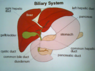
Cholestasis is a condition where bile can’t flow from the liver to the duodenum. Bile formation is a secretary function of the liver. It begins in bile Canaliculi that form between two adjacent surfaces of liver cells similar to the terminal branches of a tree. The Canaliculi join each other to form larger structures, sometimes referred to as canals of hering, which themselves join to form small bile ductules that have an epithelial surface. The ductules join to form bile ducts that eventually form either the right main hepatic duct that drains the right lobe of the liver and the left main hepatic duct draining the left lobe of the liver. The two ducts join to form the common hepatic duct, which in turn joins the cystic duct from the gall bladder, to give the common bile duct. This duct then enters the duodenum at the ampulla of Vater.       

Causes: - The causes of cholestasis are divided into two groups:
i.)                Those originating within the liver and
ii.)              Those originating outside the liver. 

Within The Liver: - Causes include:
i.)         Acute Hepatitis,
ii.)       Alcoholic Liver Disease,
iii.)      Primary biliary Cirrhosis with inflammation and scarring of the         
            bile ducts,                  
iv.)      Cirrhosis due to viral hepatitis B or C,
v.)       Drugs,
vi.)      Hormonal effects on bile flow during Pregnancy and
vii.)     Cancer that has spread to the liver.

Outside The Liver: - Causes include:
i.)                A stone in a bile duct, narrowing of a bile duct,
ii.)              Cancer of a bile duct,
iii.)           Cancer of the Pancreas and
iv.)            Inflammation of the Pancreas.

Symptoms: -
i.)                Jaundice, dark urine, light colored stools and generalized itchiness are characteristic Symptoms of Cholestasis.
ii.)              Jaundice results from excess bilirubin deposited in the skin, and dark urine results from excess bilirubin excreted by the kidneys.
iii.)           Retention of bile products in the skin may cause itching, with subsequent scratching and skin damage.
iv.)            Stools may become light-colored because the passage of bilirubin into the intestine in blocked.
v.)              Stools may contain too much fat because bile can’t enter the intestine to help digest fat in foods.
vi.)            Fatty stools may be foul-smelling. The lack of bile in the intestine also means that calcium and vitamin D are poorly absorbed. If Cholestasis Persists, a deficiency of these nutrients can cause loss of bone tissue. Vitamin K, which is needed for blood clotting, is also poorly absorbed from the intestine, causing a tendency to bleed easily.
vii.)         Prolonged jaundice due to cholestasis produces a muddy skin color and fatty yellow deposits in the skin. Whether the person has other symptoms, such as abdominal pain, loss of appetite, vomiting or fever depends on the cause of cholestasis.
Diagnosis
A doctor tries to determine whether the cause is within or outside the liver on the basis of symptoms and the results of a physical examination. Recent use of drugs that can cause cholestasis suggests a cause within the liver. Small spiders like blood vessels visible in the skin, an enlarged spleen and fluid in the abdominal cavity, which are signs of chronic liver diseases also suggest a cause within the liver.
Findings that suggest a cause outside the liver include certain kinds of abdominal pain and an enlarged gall bladder.

Laboratory Test: - The blood levels of two enzymes alkaline phosphatare and gammaglutamyl transpeptidase are very high in people with cholestasis.

Treatment
Homeopathic Medicine: - Berberis V., Carduus Mar, Chelidonium, China, Hydrastis, Lachesis, Leptandra, Lycopodium, Merc. Sol., Myrica, etc.

Saturday, 14 September 2013

Asthma

ASTHMA

Asthma is a chronic disease of the respiratory system in which the airway occasionally constricts, becomes inflamed, and is lined with excessive amounts of mucus, often in response to one or more triggers.

Pathophysiology
Inflamed airways and bronchoconstriction in asthma, airways narrowed as a result of the inflammatory response cause wheezing.

Bronchoconstriction
During an asthma episode, inflamed airways react to environmental triggers such as smoke, dust, or pollen. The airways narrow and produce excess mucus, making it difficult to breathe. In essence, asthma is the result of an immune response in the bronchial airways.
The airways of asthmatics are “hypersensitive” to certain triggers, also known as stimuli. In response to exposure to these triggers, the bronchi contract into spasm. Inflammation soon follows, leading to a further narrowing of the airways and excessive mucus production, which leads to coughing and other breathing difficulties.

Diagnosis
A Physician can diagnose asthma on the basis of typical findings in a patient’s clinical history and examination. Asthma is strongly suspected if a patient suffers from eczema or other allergic conditions-suggestion a general Atopic Constitution-or has a family history of asthma.
Testing Peak Flow at rest and after exercise can be helpful, especially in young asthmatics that may experience only exercise-include asthma
If the diagnosis is in doubt, a more formal Lung Function Test may be conducted.
Once a diagnosis of asthma is made, a patient can use peak flow meter testing to monitor the severity of the disease.
A Spirometry Test can also measure how well your lungs are functioning but provides more detailed information than peak flow meter.
Less commonly, other tests such as Chest X-rays are done to make sure do not have any other breathing problems. For example; a disease called chronic obstructive pulmonary disease has similar symptoms to asthma.
An Allergy Skin Test may be done to find out whether you are allergic to certain substances.

Signs and Symptom
Some or all of the following symptoms may be present in those with asthma:
i.)                 Dyspnea,
ii.)               Wheezing
iii.)             Stridor
iv.)            Coughing,
v.)              An inability for physical exertion.
vi.)            Some asthmatics that have severe shortness of breath and tightening of the lungs never wheeze or have Stridor and their symptoms may be confused with a COPD-type disease.

Treatment
Asthma cannot be cured. Treatment will aim to reduce the frequency, severity and length of your asthma attacks. Because a lot of different factors are involved in asthma, your treatment plan will be individual to a patient’ itself, combining medicines and asthma management in the way that works best for the patient.

Homeopathic Medicines
Aconite, Bryonia, Arsenic, Blatta orientalis, Boerhaavia diffusa, Calotropis gigantean, Justicia adhatoda, Ocimum sanctum, Gridelia, Ipecacuanha, Passiflora incarnate, Eucalyptus, Lemna Minor, etc.

Sinusitis

SINUSITIS

Sinusitis is an inflammation of the paranasal sinuses, which may or may not be as a result of infection, from bacterial, fungal, viral, allergic issues.
Newer classifications of sinusitis refer to it as Rhinosinusitis, taking into account the thought that inflammation of the nose as well as Rhinitis.

There are several paired paranasal sinuses, including the Frontal, Ethmoid, Maxillary and sphenoid sinuses.

Acute Sinusitis
 Acute Sinusitis is usually precipitated by an earlier upper respiratory tract infection, generally of viral origin, virally damaged surface tissues are then colonized by bacteria, most commonly Haemophilus Influenza, Streptococcus Pneumoniae, moraxella Cutarrhalis and Staphylococcus aureus.
Another possible cause of Sinusitis can be dental problem that affect the maxillary Sinus.
By Duration- going on less than four weeks.

Chronic Sinusitis
 Chromic Sinusitis is a complicated spectrum of disease that shares Chronic Inflammation of the Sinus.
   The Causes are multifactorial and many include allergy, environmental factors such as dust or pollution, bacterial infection and fungus. Non allergic factors such as Vasomotors Rhinitis can also cause Chronic Sinus Problems.

Diagnosis
Asymmetric growth of tongue plaque happens due to drainage from a fungal sinus infection.
  Allergies, structural problems such as for example a deviated spectrum, small sinus Ostia, smoking, nasal polyps, carrying the cystic fibrosis gene, Priorbouts of Sinusitis as each instance may result in increased inflammation of the nasal or sinus mucosa and potentially further narrow the openings.
Acute Sinusitis often last longer and typically causes more Symptoms than just a cold.
A doctor can diagnose Sinusitis by medical history, Physical examination, X-rays and of necessary MRTS or CT scan.

Treatment
There are many remedies that react well in our homeopathic system. Let us discuss indications of few remedies that follow,
Belladonna, Sanguirium Nitricum, Sanguinaria, Usnea Barbata, Prunus Spinosa, Natrum Mur, Silicea, Allium Cepa, Lemna Minor and Spigelia.

Saturday, 31 August 2013

JAUNDICE Disease

JAUNDICE

Jaundice, also known as Icterus, is a yellowing of the skin, Conjunctiva and mucous membranes caused by increased level of bilirubin in RBC (Red Blood Cells). Usually the concentration of bilirubin in the blood must not exceed 2-3 Mg/dl for the colouration to be visible. Jaundice typically appears in a ‘Top To Bottom’ Progression, and resolves in a ‘Bottom to Top’ manner.
There are Three Different Classes of Causes for Jaundices:-
i.)                  Pre-Hepatic
ii.)         Hepatic
      iii.)        Post- Hepatic
Pre- Hepatic
Pre-Hepatic Jaundice is caused by anything which causes an increased rate of Hemolysis. In tropical Countries, Malaria can cause Jaundice in this manner. Certain Genetic diseases, Such as sickle cell Anemia and Glucose6- Phosphate Dehydrogenase deficiency can lead to increased red cell lysis and therefore Hemdytic Jaundice. Defeets in bilirubin metabolism also present as Jaundice. Jaundice usually comes with high fevers.
HEPATIC 
Hepatic causes include acute hepatitis, hepatoxicity and alcoholic liver disease, whereby cell necrosis reduces the liver’s ability to metabolise and excrete bilirubin leading to a buildup in the blood. Less common causes include primary biliary Cirhosis, Gilbert’s Syndrome and metastatic Careinoma. Jaundice seen in the newborn, known as Neonatal Jaundice, is common, occurring in almost every newborn as Hepatic machinery for the conjugation and excretion of bilirubin does not fully mature until approximately two weeks of age.
Post-Hepatic
Post-Hepatic Jaundice, also called cholestasis, is caused by an interruption to the drainage of bile in the biliary system. The most common causes are Gallstones in the common bile duct and Panaoreatic Cancer in the head of the Pancreas, also a group of Parasites known as ‘Liverflukes’ live in the common bile duct, ductal Carcinoma, Pancreatitis and Pancreatic Pseudocysts. A rare cause of obstructive Jaundice is Mirizzi’s Syndrome.
The Presence of Pale Stools and dark urine suggests an Obstructive or Post-Hepatic cause as normal faces get their color from bile pigments. Patients often complain of severe Itching.

Diagnosis and Treatment   
A doctor uses laboratory tests and imaging studies to determine the cause of the Jaundice.
There are many remedies that react well in our Homeopathic System. Let us discuss Indications of few remedies that follow.

Carduus Marianus, Ceanothus Americarius, Chelielonium Majus, Cinchona Off, Dolichos Pruriens, Kalmegh, Nux-Moschates, etc.

If this post helps you than please share your experience with us.....

Liver Disease

LIVER

The Liver is located in the upper right- hand portion of the abdominal cavity, beneath the diaphragram and on top of the
Stomach, right kidney and Intestines shaped like a triangle, the lever is a dark raddish-brown organ that weighs about three Pounds.

There are two distinct sources that supply blood to the liver:-

        i.) Oxygenated blood flows in from the hapatic artery.

      ii.) The portal veins brings nutritious venous blood from the Intestine & spleen.

The liver holds about one Pint (13%) of the body's blood supply at any given moment.

 LIVER DISORDER

Sometimes the manifestations of liver disease are not obvious. For example, Symptons may include fatigue, a feeling of 
unwellness, loss of appetite and mild weight loss. However, there symptons are also typical of many other diseases. Thus,
liver disease can easily be overlooked, Particularly in its early stages.

Some major Features of Liver Disease:-
Jaundice:-      
 A yellowish discoloration of the skin and white of an eyes (Sclera).

Hepatomegaly:-          
                                                           Liver enlargement.

Ascites:-         
                                                Fluid in the abdominal Cavity.
   
Hepatic:-              
     Confusion Caused by deterioration of brain function due to buildup of toxic substances in 
                                                                    the blood.

Gastrointestinal bleeding:-
 Bleeding from large, tortous Veins in the esophagus and stomach.

Portal Hypertension:-       
Aboriormally high blood pressure in the veins that bring blood from the Intestine to the Liver.

SKin Symptons:-            
 Spider like blood vessels on the face and chest & Red Palms. Brightred 
Complexion Itching.

Blood Abnormalities:-      
 Decreased number of red blood cells (anaemia). Decreased number of white cells (Leukopenia). 
                            Decreased number of platelets (Thrombocytopenia).A tendency to bleed (Coagulopathy).

Hormonal Abnormalities:- 
   High levels of Insulin
  HUMLIN.
 NOVOLIN.
   But a poor response to it, leading to high blood sugar levels.
                            Cessation of menstrual Periods and decreased fertility in women. Erectile dysfunction and feminization in men.
            
Heart And Blood Vessels Abnormalities:- 
  Increased heart rate and amount of blood pumped. Low blood Pressure.

General Symptons:-          
Fatigue, Weakness, Weight Loss, Poor Appetite Nausea, Fever, Abdominal Pain.

If this post helps you than please share your experience with us.....

 
Copyright @ 2013 Homeopath.